Most people living with keratoconus have heard of cross-linking, scleral lenses, and corneal transplants. CTAK, or Corneal Tissue Addition Keratoplasty, doesn’t come up nearly as often, partly because it’s only been available for a few years and partly because not every cornea specialist performs it yet. This newer procedure is quietly giving patients with advanced keratoconus a way to improve vision and reshape the cornea without taking the bigger step of a full transplant.
What Is Keratoconus?
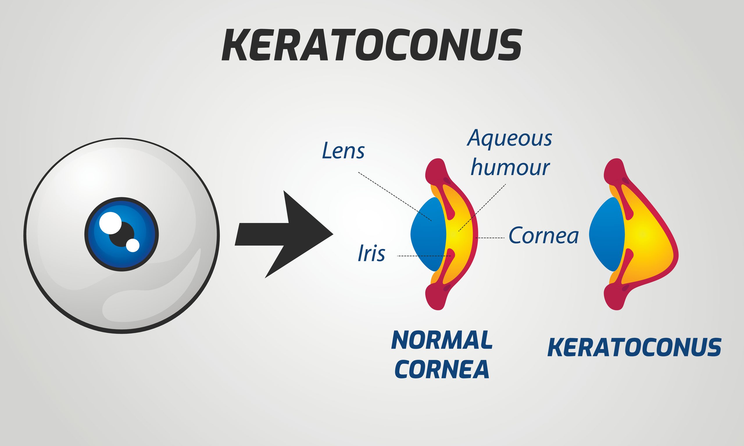
The cornea is the clear, dome-shaped front surface of the eye, and its job is to bend light accurately onto the retina. In a healthy eye, the cornea holds a relatively smooth, rounded shape. In keratoconus, the structural integrity of the cornea gradually weakens, causing it to thin and bulge outward into an irregular cone shape. That distortion scatters incoming light, producing blurred and distorted vision that standard eyewear often cannot fully correct.
Keratoconus typically develops in the teens and twenties and tends to progress through the mid-thirties or so, though the rate varies considerably from person to person. In its early stages, updated glasses or specially fitted contact lenses can often compensate for the irregular astigmatism keratoconus creates. As the condition progresses, those compensations become less effective.
The Treatment Gap CTAK Fills
The treatment ladder for keratoconus has traditionally had a significant gap in the middle. At one end, corneal cross-linking is the standard intervention for progressive keratoconus. It uses ultraviolet light and riboflavin eye drops to stiffen corneal fibers and slow or halt progression, but it does not restore the corneal shape that has already been lost. At the other end, corneal transplant surgery replaces the central cornea entirely, but it carries significant recovery time, surgical risk, and a long period of vision rehabilitation afterward.
That leaves patients with moderate to advanced keratoconus, who have moved past what cross-linking alone can address but who are not yet ready, or willing, to undergo a transplant. For years, this group had few surgical options. CTAK was developed specifically to address that gap.
What Is CTAK?
Rather than removing or replacing corneal tissue, the CTAK procedure adds donor tissue to reshape the cornea from within. A thin, arc-shaped inlay made from preserved, gamma-irradiated donor corneal tissue is implanted into a precisely created channel inside the patient’s corneal stroma. The added tissue flattens the irregular cone and restores a more normal curvature, which improves how the eye focuses light.
What distinguishes CTAK from older approaches is the degree of customization. Each inlay’s size, shape, and placement are customized to the patient’s corneal topography.
The surgeon receives both the customized inlay and a suggested surgical plan based on the patient’s imaging data. This level of precision is what allows CTAK to produce significant improvements across a wide range of keratoconus presentations, from mild to more advanced cases.
Because the procedure adds tissue rather than removing it, the patient’s native corneal structure remains largely intact. This is an important distinction from corneal surgery approaches that involve more extensive tissue replacement, and it is one reason CTAK can delay or prevent the need for a full-thickness transplant.
How Does the CTAK Procedure Work?
The process begins well before the day of surgery. During your consultation, a detailed corneal mapping using topography and tomography captures the shape, thickness, and characteristics of the patient’s cornea. That data is used to design the custom inlay, which is then processed and shipped to the surgeon before the procedure.
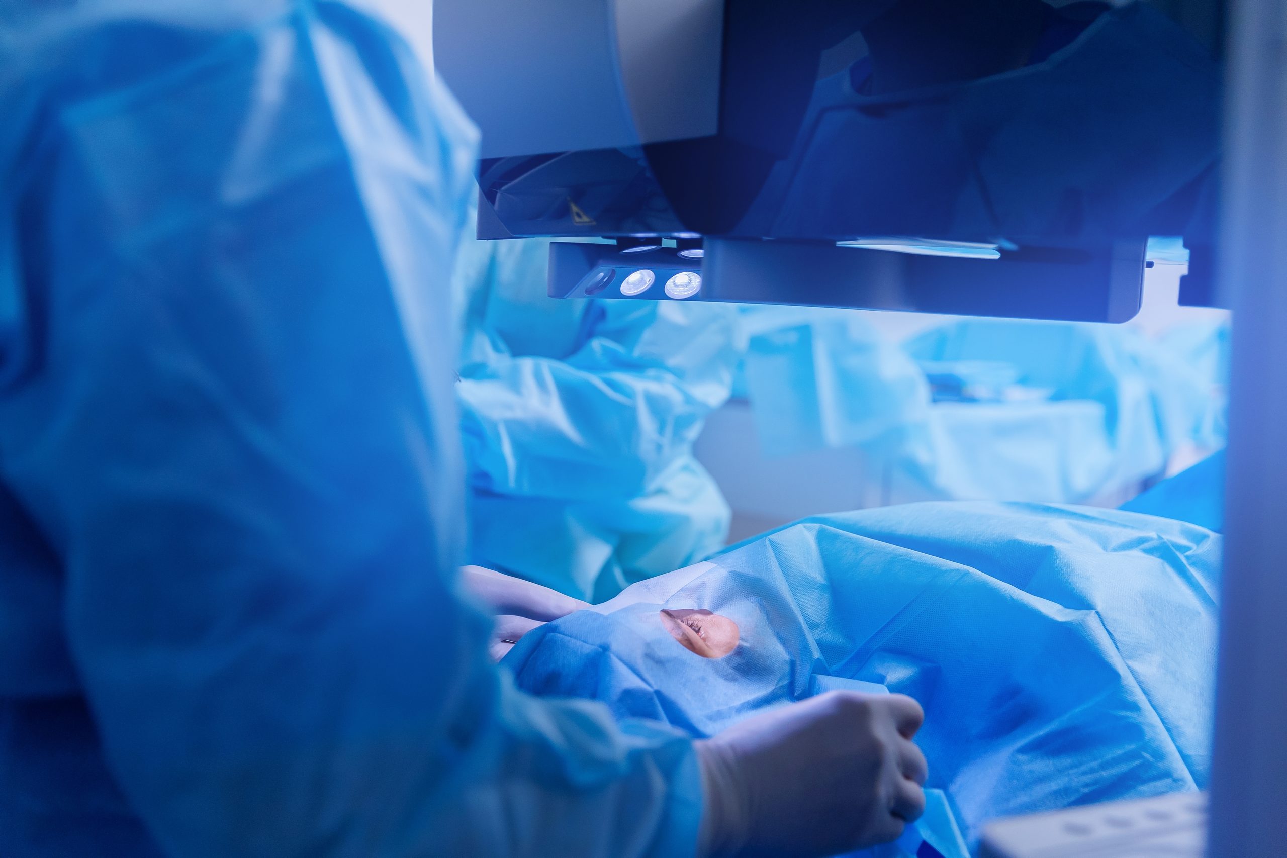
On the day of surgery, the patient’s eye is numbed with anesthetic drops. The surgeon uses a femtosecond laser to create a precise channel within the corneal stroma, sized and positioned to match the planned inlay. The donor tissue inlay is then inserted through a micro-incision and guided into the channel. Once positioned correctly, it is smoothed into place. In most cases, no sutures are required. The entire procedure is performed on an outpatient basis and typically takes less than an hour.
Two appointments are generally involved in the full process: one for the laser portion and a second, usually within two weeks, for the tissue implantation itself.
Most patients return home the same day, with a protective contact lens placed on the eye to support healing. Mild irritation and light sensitivity are common during the first few days, and prescription drops are used to prevent infection and inflammation. Many patients notice visual improvement within the first week, with results continuing to refine over the following months.
Who Is a Good Candidate for CTAK?

CTAK works best for patients whose keratoconus falls within a moderate to advanced range. Patients with very peripheral steepening or significant central corneal scarring that limits visual potential may not be ideal candidates.
One patient population that deserves particular attention is those who have been told a corneal transplant is their only remaining surgical option. For many of these patients, CTAK may provide a meaningful alternative worth discussing. The same applies to patients who have developed corneal ectasia following refractive surgery, where CTAK can help stabilize and reshape weakened tissue.
Candidacy is always determined through a thorough consultation and corneal imaging, which provides the full picture needed to evaluate whether CTAK is the right approach.
Has your keratoconus progressed beyond what glasses or contacts can correct? Schedule an appointment at RGB Cataract & LASIK in Sherman, TX.